Scientific Manuscripts and Original Research

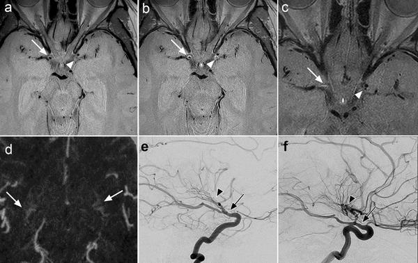
Vessel wall imaging features of Moyamoya disease in a North American population: patterns of negative remodelling, contrast enhancement, wall thickening, and stenosis

This study characterized vessel wall imaging (VWI) features of Moyamoya disease (MMD) in a predominantly adult population at a North American center.MethodsConsecutive patients with VWI were included. Twelve arterial segments were analyzed for wall thickening, degree and pattern of contrast enhancement, and remodeling.ResultsOverall, 286 segments were evaluated in 24 patients (mean age = 36.0 years [range = 1–58]). Of 172 affected segments, 163 (95%) demonstrated negative remod...

The Frequency of Carotid Intraplaque Hemorrhage on Vessel... : Journal of Neuro-Ophthalmology

Central retinal artery occlusion (CRAO) and branch retinal artery occlusion (BRAO) are analogous to an acute stroke of the eye and represent a neurological emergency because they may be a harbinger of a central nervous system stroke (1–3). CRAO and BRAO (collectively referred to as retinal artery occlusion, RAO, henceforth) commonly occur as a result of an embolic event in the context of carotid artery plaque (4). Various features of carotid plaque increase the risk for ischemic cerebral events...
Photo by cottonbro studio on Pexels

Normalized intraplaque hemorrhage signal on MP-RAGE as a marker for acute ischemic neurological events

This study sought to validate whether the signal intensity ratio (SIR) of carotid intraplaque hemorrhage (IPH) was associated with acute ischemic neurologic events. A retrospective review was completed of consecutive patients that underwent neck magnetic resonance angiography using magnetization prepared-rapid gradient echo (MP-RAGE) and T1-CUBE sequences between 2017 and 2020. Patients with magnetic resonance evidence of IPH were included. SIRs were measured by comparing the maximum IPH signal with the mean intramuscular signal from the adjacent sternocleidomastoid. Patients were stratified into ischemic or non-ischemic groups based on the presence of acute ipsilateral ischemic events (stroke, retinal artery occlusion). Logistic regression analysis was performed to determine if increasing IPH SIR was associated with an increased risk of ipsilateral ischemic events. Of 85 included patients (85 arteries), 66 were male (77.6%). Mean age was 71.0 (SD ± 11.1). There were 70 arteries with IPH that were ipsilateral to an ischemic event, and 15 that belonged to patients without an ischemic event. No association was found between increasing IPH SIR seen on MP-RAGE (odds ratio (OR): 0.82; 95% confidence interval (CI): 0.58–1.4; P = 0.43) or T1-CUBE sequences (OR: 0.85; 95% CI: 0.53–1.5; P = 0.56). There was no association between the SIR of IPH and acute ischemia on either MP-RAGE or T1-CUBE sequences. Further investigation is required prior to widespread acceptance of SIR as a predictive imaging marker of symptomatic carotid plaque.

Carotid Intraplaque Hemorrhage and Stenosis: At What Stage of Plaque Progression Does Intraplaque Hemorrhage Occur, and When is It Most Likely to Be Associated with Symptoms?

The relationship between carotid intraplaque hemorrhage and luminal stenosis severity is not well-established. We sought to determine whether intraplaque hemorrhage is related to carotid stenosis and at what degree of stenosis intraplaque hemorrhage most likely contributes to ischemic symptoms. Consecutive patients who underwent MR carotid plaque imaging with MPRAGE sequences to identify intraplaque hemorrhage were retrospectively reviewed. Degrees of stenoses were categorized as minimal (<30%), moderate (30%–69%), and severe (>70%). Arteries were categorized into 2 groups: symptomatic (ipsilateral to a cerebral ischemic event) and asymptomatic (from a patient without an ischemic event). Multiple regression analyses were used to determine independent associations between the degree of stenosis and intraplaque hemorrhage and the presence of intraplaque hemorrhage with symptoms among categories of stenosis. We included 449 patients with 449 carotid arteries: Two hundred twenty-five (50.1%) were symptomatic, and 224 (49.9%) were asymptomatic. An increasing degree of stenosis was independently associated with the presence of intraplaque hemorrhage (OR = 1.02; 95% confidence interval, 1.01–1.03). Intraplaque hemorrhage was independently associated with ischemic events in arteries with <30% stenosis (OR = 5.68; 95% CI, 1.49–21.69). No such association was observed in arteries with >30% stenosis. Of symptomatic arteries with minimal stenosis, 8.7% had intraplaque hemorrhage versus 1.7% of asymptomatic arteries (P = .02). No differences in intraplaque hemorrhage prevalence were found between symptomatic and asymptomatic groups with moderate (P = .18) and severe stenoses (P = .99). The presence of intraplaque hemorrhage on high-resolution plaque imaging is likely most useful in identifying symptomatic plaques in cases of minimal stenosis.

Embolic Stroke of Undetermined Source: The Association With Carotid Intraplaque Hemorrhage

Embolic stroke of undetermined source (ESUS) is defined as a brain infarct without a hemodynamically significant proximal artery stenosis or high-risk cardioembolic source such as atrial fibrillation. In addition to the degree of stenosis, advancements in imaging technology have enabled stratification for the future risk of stroke based on the presence of high-risk plaque features including intraplaque hemorrhage (IPH). IPH likely represents an under-recognized source of thromboembolism among patients with ESUS. We sought to further explore this potential relationship by assessing the presence of IPH among patients with embolic stroke or transient ischemic attack of undetermined source and correlating IPH with the degree of stenosis ipsilateral to the ischemic event.

Reductions in bilateral transverse sinus pressure gradients with unilateral transverse venous sinus stenting for idiopathic intracranial hypertension

Venous sinus stenting is an effective treatment for papilledema associated with idiopathic intracranial hypertension (IIH). It is unclear whether unilateral transverse-sigmoid sinus (TSS) stenting adequately decompresses the contralateral TSS system in cases of bilateral transverse sinus stenosis. The objective of this study was to compare changes in bilateral TSS pressure gradients following unilateral TSS stenting in a series of patients with IIH. Consecutive patients from a...
Photo by Anna Shvets on Pexels

Variations in the Presence of Carotid Intraplaque Hemorrhage Across Age Categories: What Age Groups Are Most Likely to Benefit From Plaque Imaging?

Although carotid artery intraplaque hemorrhage (IPH) is a known risk-factor for cerebral ischemic events in patients of advanced age, its prevalence in younger cohorts is less certain. The purpose of this study was to assess the prevalence of carotid artery IPH across the age spectrum. A retrospective review was completed of all adult patients from our institution who underwent neck MRA with high-resolution carotid plaque imaging between 2017 and 2020. The mean ages of patients with and without...
Photo by DS stories on Pexels

Left-sided carotid arteries have a higher prevalence of intraplaque hemorrhage than right-sided: An asymmetric conundrum

To assess whether an asymmetry exists in the prevalence of carotid artery intraplaque hemorrhage (IPH) between right- and left-sided arteries. The records of all patients with atherosclerotic carotid artery disease that underwent neck magnetic resonance angiography imaging with high-resolution plaque sequences between 2017 and 2020 at our institution were retrospectively reviewed. The prevalence of stenosis and IPH was determined for all patients and compared between the left and right carotid arteries of those with unilateral anterior circulation ischemic strokes. Multiple regression analysis was performed to determine potential independent associations of IPH laterality with ischemic strokes. A total of 368 patients were included overall and 241 were male (65.4%). There were a total of 125 asymptomatic patients and 211 patients with unilateral anterior circulation ischemic strokes. Of patients with ischemic strokes, 55.5% had left-sided strokes compared with 44.5% who had right-sided strokes (p = 0.03). Patients with left-sided strokes had a higher prevalence of ipsilateral IPH than those with right-sided strokes (64.1% versus 36.2%, p < 0.0001), despite similar degrees of stenosis. Both age (odds ratio (OR): 1.0; 95% confidence interval (CI): 1.0–1.1; p = 0.007) and the presence of left-sided IPH (OR: 3.2; 95% CI: 1.5–6.8; p = 0.003) were independently associated with unilateral ischemic strokes. Left-sided plaques more frequently have IPH and may be more likely to result in ipsilateral ischemic strokes compared with right-sided plaques. The underlying mechanism of asymmetric distribution of IPH between right and left carotids remains unclear.

Variations of Intracranial Dural Venous Sinus Diameters from Birth to 20 Years of Age: An MRV-Based Study

The role of the dural venous sinus system in cerebrovascular pathology and the understanding of normal developmental patterns and sizes of the dural venous sinus system continue to expand. The purpose of this study was to review MR venograms to elucidate developmental patterns and diameters of the major dural venous sinuses from 0 to 20 years of age. All available MR venograms of patients 0–20  years of age who presented to our institution were retrospectively reviewed. Patient age at the time of image acquisition was noted, and measurements were taken of the diameters of the major dural venous sinuses. The presence of embryonic sinuses including the persistent falcine sinus and the occipital sinus was noted. Dominance patterns of the transverse sinus system were determined. Mean diameters of each sinus were plotted as a function of age. The prevalence of persistent prenatal sinuses and transverse sinus–dominance patterns was compared across ages. A total of 429 MR venograms from 429 patients were reviewed. All dural venous sinuses demonstrated a maximal growth rate from 0 to 7 years of age and reached maximal diameters around 5–10 years of age. The prevalence of falcine sinuses and occipital sinuses trended downward across increasing age categories (P = .09 and, <.0001, respectively). Dural venous sinuses demonstrate maximal growth between 0 and 7 years of age and reach adult size around 5–10 years of age. Involution of the prenatal sinuses continues to take place after birth into childhood but is largely absent in early adulthood.
Photo by MART PRODUCTION on Pexels

Intracranial Aneurysms in White Patients with Moyamoya Disease: A U.S. Single-Center Case Series and Review

Because of the changes in hemodynamics and the structure of the arterial wall, cerebral arteries in patients with MMD may also form intracranial aneurysms (IAs). In patients with MMD, the prevalence of IAs may be as high as 18% compared with 2%–3% in the general population. In addition, the characteristics of MMD, including narrow internal carotid arteries (ICAs), fragile arterial walls, high density of basilar collateral vessels, and so on, make the treatment of IAs in patients with MMD p...

High prevalence of pro-thrombotic conditions in adult patients with moyamoya disease and moyamoya syndrome: a single center study - Acta Neurochirurgica

Moyamoya Disease (MMD) and moyamoya Syndrome (MMS) have been reported to be associated with pro-thrombotic states in some patients. To date, however, such reports have been limited to case reports or small case series. We sought to determine the prevalence of pro-thrombotic states among a large cohort of both MMD and MMS patients.We retrospectively reviewed the medical records of all adult patients who were diagnosed with MMD or MMS from our institution. In addition to basic demographic and clin...