Review Articles

Emerging Roles of Exosomes in Stroke Therapy

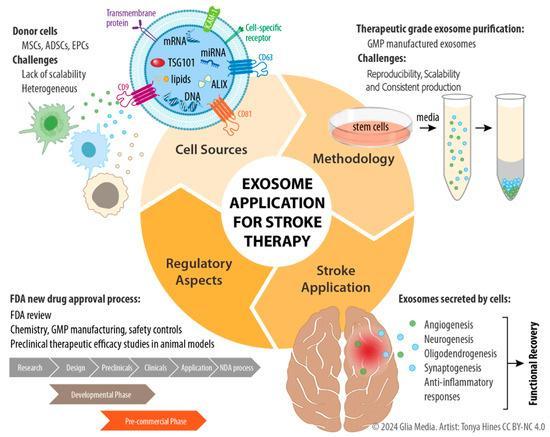
Stroke is the number one cause of morbidity in the United States and number two cause of death worldwide. There is a critical unmet medical need for more effective treatments of ischemic stroke, and this need is increasing with the shift in demographics to an older population. Recently, several studies have reported the therapeutic potential of stem cell-derived exosomes as new candidates for cell-free treatment in stoke. This review focuses on the use of stem cell-derived exosomes as a potential treatment tool for stroke patients. Therapy using exosomes can have a clear clinical advantage over stem cell transplantation in terms of safety, cost, and convenience, as well as reducing bench-to-bed latency due to fewer regulatory milestones. In this review article, we focus on (1) the therapeutic potential of exosomes in stroke treatment, (2) the optimization process of upstream and downstream production, and (3) preclinical application in a stroke animal model. Finally, we discuss the limitations and challenges faced by exosome therapy in future clinical applications.
Photo by cottonbro studio on Pexels

A review of histopathologic and radiologic features of non-atherosclerotic pathologies of the extracranial carotid arteries

Diseases of the carotid arteries can be classified into different categories based on their origin. Atherosclerotic carotid disease remains the most encountered arterial wall pathology. However, other less-common non-atherosclerotic diseases can have detrimental clinical consequences if not appropriately recognized. The underlying histological features of each disease process may result in imaging findings that possess features that are obvious of the disease. However, some carotid disease processes may have histological characteristics that manifest as non-specific radiologic findings. The purpose of this manuscript is to review various non-atherosclerotic causes of carotid artery disease as well as their histologic-radiologic characteristics to aid in the appropriate recognition of these less-commonly encountered pathologies.

Nonstenotic Carotid Plaques and Embolic Stroke of Undetermined Source: A Multimodality Review

Symptomatic nonstenotic carotid artery disease has been increasingly recognized as a thromboembolic source in patients who would otherwise be classified as having embolic stroke of undetermined source. Evidence suggests that certain plaque features seen on sonography, CT, and MR imaging in nonstenotic carotid artery disease may predispose to recurrent stroke in patients with embolic stroke of undetermined source. We performed a focused literature review to further study plaque features in the context of embolic stroke of undetermined source and to determine which plaque features may be associated with ipsilateral ischemic events in such patients. Plaque thickness as seen on both ultrasound and CT appears to have a consistent association with ipsilateral stroke in patients with embolic stroke of undetermined source across multiple studies. Intraplaque hemorrhage as seen on MR imaging is now understood to have a strong association with ipsilateral stroke in patients with embolic stroke of undetermined source. Continued study of various plaque features as seen on different modalities is warranted to uncover other potential associations.

Endarterectomy for symptomatic non-stenotic carotids: a systematic review and descriptive analysis

Atherosclerotic disease of the carotid arteries carries a well-known association with ipsilateral cerebral ischaemic events, including stroke. As a result of landmark trials including the North American Symptomatic Carotid Endarterectomy Trial (NASCET), decisions regarding invasive treatment have been based primarily on the degree of carotid luminal stenosis. Indeed, the current guidelines reflect the central role of the degree of stenosis in determining whether intervention is war...
Photo by meo on Pexels

The cerebrofacial metameric syndromes: An embryological review and proposal of a novel classification scheme

Cerebrofacial metameric syndromes (CMS) are congenital disorders caused by embryological defects in craniofacial vasculogenesis, which results in vascular malformations within predictable anatomical patterns of the brain, cranium and face.
Syndromic presentation can manifest with either high-flow or low-flow malformations, termed cerebrofacial arteriovenous metameric syndrome (CAMS) or cerebrofacial venous metameric syndrome (CVMS), respectively. High-flow vascular malformations in CAMS are m...
Photo by MART PRODUCTION on Pexels

Imaging of benign cervicofacial vascular anomalies and associated syndromes

Cervicofacial vascular anomalies can result in morbidity, pain, and cosmetic concerns in affected individuals. Each anomaly has its own unique natural history, treatment, and associations with underlying genetic syndromes. For optimal patient care, it is important for the neuroradiologist to accurately recognize and characterize these entities to ensure appropriate treatment and management. In this review, we discuss the general characteristics, classifications, and imaging features associated with the most common vascular anomalies such as hemangiomas, arteriovenous malformations and fistulas, capillary malformations, venous malformations, and lymphatic malformations in the context of associated syndromes. Additionally, we discuss novel imaging techniques that aid in identifying these vascular anomalies.
Photo by cottonbro studio on Pexels

Minimally-invasive tubular retraction ports for intracranial neurosurgery: History and future perspectives

Brain retraction is a necessary yet potentially damaging requirement of accessing lesions located in deep structures. The development of minimally-invasive tubular retractors (MITRs) provides the theoretical advantage of maximizing visualization of and access to deep-seated lesions, all while minimizing collateral tissue damage. These advantages make MITRs preferable to traditional bladed retractors in the majority of deep-seated lesions. Several commercially-available MITR systems currently exist and have been shown to aid in achieving excellent outcomes with acceptable safety profiles. Nevertheless, important drawbacks to currently-available MITR systems exist. Continued pursuit of an ideal MITR system that provides maximal visualization and access to deep-seated lesions while minimizing retraction-related tissue damage is therefore important. In this review, we discuss the historical development of MITRs, the advantages of MITRs compared to traditional bladed retractors, and opportunities to improve the development of prospective MITRs.

Implementation and Rationale for a Unified Clinical and Imaging Protocol for Evaluation and Treatment of Moyamoya Angiopathy: A Single Institutional Experience

Moyamoya disease (MMD) is a complex and incompletely-understood cerebrovascular pathological entity that requires thorough clinical and imaging evaluation. Moyamoya is rare, thereby making the establishment of an effective, thorough and interdisciplinary patient evaluation protocol challenging, even within specialized referral centers. Nevertheless, implementation of such a protocol is crucial in order to provide the best possible evaluation and treatment for MMD patients. Here, we describe our...
Photo by MART PRODUCTION on Pexels

Subarachnoid Hemorrhage of Unknown Cause: Distribution and Role of Imaging

•Subarachnoid hemorrhage of unknown cause is a fairly common occurrence and comprises 10% to 15% of all nontraumatic subarachnoid hemorrhages.

•Such cases can be categorized into distinct groups based on the location and distribution of subarachnoid blood.

•Each group of subarachnoid hemorrhage of unknown cause carries unique imaging recommendations in acute and follow-up contexts.

Neurosurgical management of aneurysms of the vertebrobasilar system: increasing indications for endovascular therapy with a continued role for open microneurosurgery

Aneurysms of the vertebrobasilar system remain among the most challenging subsets of aneurysms to treat with an open surgical approach. Since Charles Drake’s pioneering work in the 1960s, several advances in microsurgical techniques have improved outcomes and feasibility in the open surgical management of these aneurysms. In parallel, the field of endovascular neurosurgery has provided several safe and effective treatment options. Multiple trials have suggested that endovascular therapy for aneurysms of the vertebrobasilar system is superior to open surgical management in most cases. In some instances, however, open surgical management likely represents a more effective and durable option relative to endovascular therapy. Therefore, continued training of future cerebrovascular specialists in open surgery of vertebrobasilar aneurysms remains crucial. With widespread utilization of endovascular techniques, however, proper exposure of trainees to such aneurysms is growing increasingly difficult. In this review, we discuss the recent advances in the endovascular management of vertebrobasilar aneurysms while also emphasizing the continued importance of open microneurosurgery in such cases.

Lack of Baseline Intracranial Aneurysm Wall Enhancement Predicts Future Stability: A Systematic Review and Meta-Analysis of Longitudinal Studies

BACKGROUND: The utility of vessel wall MR imaging in identifying unstable intracranial aneurysms has been suggested but remains controversial.

PURPOSE: Our aim was to provide further insight into the potential relationship between aneurysm wall enhancement on initial vessel wall imaging and aneurysm instability at follow-up.

DATA SOURCES: Our sources were PubMed, Scopus, the Web of Science, and the Cochrane Central Register of Controlled Trials.

STUDY SELECTION: We searched for English language studies that reported the presence of vessel wall enhancement of unruptured intracranial aneurysms on baseline vessel wall imaging studies with longitudinal follow-up of aneurysm status.

DATA ANALYSIS: Aneurysms were grouped into “stable” and “unstable” groups at follow-up on the basis of growth, symptomatic manifestation, or rupture. The association of each group with aneurysm wall enhancement on initial vessel wall imaging was determined.

DATA SYNTHESIS: Three studies constituting 407 aneurysms were included. Aneurysms with wall enhancement were at higher risk of being unstable at follow-up (risk ratio = 3.6; 95% confidence interval, 1.7–7.5). The sensitivity of aneurysm wall enhancement on vessel wall imaging was 74.3% (95% CI, 56.7%–87.5%), specificity was 58.3% (95% CI, 53.1%–63.4%), positive predictive value was 14.4% (95% CI, 11.8%–17.4%), negative predictive value was 96.0% (95% CI, 93.2%–97.7%), and the overall accuracy of the test was 59.7% (95% CI, 54.8%–64.5%).

LIMITATIONS: Only 3 studies were identified for inclusion in this analysis. More longitudinal studies of vessel wall imaging and aneurysm progression are needed.

CONCLUSIONS: The lack of wall enhancement may be a predictor of aneurysm stability. The utility of vessel wall imaging in detecting unstable aneurysms requires more data.

From the Historical Examples of Drs. Osler, Cushing, and Van Wagenen: Lessons on the Importance of Mentorship in Contemporary Neurosurgery

Although the historical relationships between William Osler, Harvey Williams Cushing, and William Perine Van Wagenen are well known in the neurosurgical world, the nature of the mentor-mentee relationships that existed between these historical giants is not widely appreciated. In this historical vignette, we describe and exemplify such relationships, while at the same time extract important and applicable principles from them. We reviewed relevant primary and secondary sources that documented the interactions between Cushing, Osler, and Van Wagenen. In founding the field of neurological surgery, the brilliant yet volatile Dr. Harvey Cushing received guidance from his mentor, Dr. William Osler. Through our review, it is undeniable that Dr. Osler's personal and professional guidance was vital to young Dr. Cushing's success as the founder of modern neurosurgery. Likewise, Cushing's tutelage of Van Wagenen enabled Van Wagenen to become a leader of a second generation of neurosurgeons, thereby perpetuating the existence of Cushing's high neurosurgical standards. These historical mentor-mentee relationships were built on 4 primary components: accurate recognition of talent, guidance, arrangement of opportunity, and sustenance of mentorship—actions that are commonly implicated in effective mentorship in contemporary studies. Proper mentorship remains indispensable for the success of neurosurgical trainees.
Photo by Yasser Hernandez on Pexels

Cardiac manifestations in a western moyamoya disease population: a single-center descriptive study and review

An embryological association between moyamoya disease (MMD) and cardiac manifestations has been proposed. Data up to this point remains anecdotal, and the prevalence of cardiac manifestations in a western MMD population is uncertain. The objective of this study was to determine the prevalence of cardiac manifestations including coronary artery disease (CAD) and congenital cardiac defects in a mostly Caucasian population of MMD patients and review prior reports of such cases. Medical records of M...
Photo by CDC on Pexels

Coronavirus Disease 2019 and the Cerebrovascular‐Cardiovascular Systems: What Do We Know So Far?

The severe acute respiratory syndrome coronavirus 2 pandemic of 2019 to 2020 has resulted in multiple hospitalizations, deaths, and economic hardships worldwide. Although respiratory involvement in patients with coronavirus disease 2019 (COVID‐19) is well known, the potential cardiovascular and cerebrovascular manifestations are less understood. We performed a PubMed and Google Scholar search and reviewed relevant literature on COVID‐19 and cardiovascular system involvement. Severe acute respiratory syndrome coronavirus 2 possesses high affinity for angiotensin‐converting enzyme 2 receptor, which is highly concentrated in the lungs and cardiovascular tissue, thereby provoking concern for cardiovascular involvement in COVID‐19 cases. Preexisting cardiovascular and cerebrovascular disease has been shown in previous reports to be a risk factor for severe infection. On the basis of our review of published studies, COVID‐19 patients may be more likely to experience acute cardiac injury, arrhythmia, coagulation defects, and acute stroke and are likely to have poorer outcomes as a result. As the COVID‐19 pandemic continues, more data about potential cardiovascular and cerebrovascular manifestations of the disease are required.

Location-based treatment of intracranial aneurysms in moyamoya disease: a systematic review and descriptive analysis

We conducted a systematic review of the literature to evaluate the efficacy of various treatment modalities for intracranial aneurysms (IA) in patients with moyamoya disease (MMD) based on anatomical location of IA. A comprehensive review of studies documenting single cases or series of MMD patients with concomitant IA was conducted. Aneurysms were classified into two primary anatomical categories: those of the Circle of Willis (CoW) and those of peripheral “moyamoya” collateral vessels. Conservative, endovascular, and open surgical treatment modalities and their outcomes between each anatomical subgroup were descriptively compared. A total of 124 studies consisting of 275 patients with 313 IA were included. Of all IA, 59.6% were located on CoW vessels, 33.7% on peripheral vessels, and 6.7% in “other” locations. Of all CoW IA, 87.2% treated with endovascular techniques had no or minimal deficit at follow-up as compared with 56.7% of those treated with open surgery. Ninety-five percent of patients with peripheral aneurysms treated with endovascular therapy had no or minimal deficit, in contrast to open surgery (69.6%). Of peripheral IA treated conservatively with or without revascularization, 65.7% had spontaneous resolution as compared with 12.0% IA of the CoW. Our results support the use of endovascular techniques for direct treatment of both CoW and peripheral IA. Aneurysms of peripheral vessels respond well to indirect treatment through surgical revascularization as opposed to CoW aneurysms. The quality of evidence is limited due to heterogeneity of included studies and IA management in MMD patients should be considered in a case-specific manne.

Brain capillary telangiectasias: from normal variants to disease

Brain capillary telangiectasias (BCTs) are small, dilated capillary networks in the brain that are most commonly asymptomatic. Though rare, symptomatic cases of BCTs have been reported, and it is therefore important to understand the nature of these lesions in order to facilitate proper recognition. Relative to other intracranial vascular malformations, updated information on the various epidemiologic, radiographic, and pathologic features of BCTs within the published literature may be inadequat...