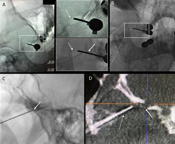
The foramen ovale “mirage” and how this impacts percutaneous cannulation for treatment of trigeminal neuralgia: a report of two cases
Percutaneous stereotactic radiofrequency rhizotomy (PSR) for trigeminal neuralgia most commonly utilizes 2D fluoroscopy for intraoperative needle guidance into the foramen ovale (FO). We describe two cases in which needle advancement into FO was unachievable despite appropriate needle placement on biplane fluoroscopy. Intraoperative multiplanar reconstruction was helpful in more accurately depicting foraminal anatomy which allowed the manipulation of the tip of the needle, which was followed by successful FO cannulation. We propose that this “mirage” is likely created by the inherent nature of X-ray-based fluoroscopy in which the FO appears to be readily penetrable, when in fact the 3D anatomy actually prevents cannulation.


